Anatomic pathology has always been the quiet engine behind modern medicine. Before a surgeon decides how aggressively to treat a tumor, before an oncologist selects a targeted therapy, before a patient is told what they are dealing with, a pathologist has examined tissue under a microscope and made a determination. That process has been central to clinical medicine for well over a century. What has changed dramatically in recent years is how that work gets done, how quickly it happens, and what tools pathologists have at their disposal when they do it.
The field is moving faster now than at any point in its history. Digital imaging, artificial intelligence, molecular diagnostics, and new laboratory management tools are reshaping what anatomic pathology looks like from the inside, and those changes are rippling outward in ways that affect every patient who ever needs a biopsy or a tissue diagnosis. Understanding what is driving this transformation, and what it actually means in practice, is worth taking the time to explore.
From Glass to Digital
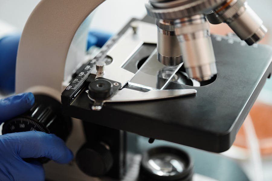
For most of its history, anatomic pathology was inseparable from the glass slide. A tissue sample would be processed, embedded in paraffin, sliced thin, stained, and mounted on glass. A pathologist would sit at a microscope, work through the slide, and render a diagnosis. The process was effective, but it had obvious limitations. Slides could be lost or damaged in transit. Getting a second opinion meant shipping physical material and waiting days for it to arrive. Reviewing large volumes of slides for quality assurance or research purposes was genuinely difficult.
Whole slide imaging has changed this. High-resolution digital scanners can now convert a glass slide into a digital image so detailed that a pathologist can zoom into cellular structures with the same precision as a physical microscope. Once digitized, a slide can be shared instantly, reviewed remotely, stored indefinitely, and accessed by multiple people at the same time. A pathologist in one city can consult with a subspecialist in another in the time it used to take to arrange a courier.
For patients in smaller communities or regions without access to specialized pathology expertise, this matters enormously. Digital pathology effectively removes geography as a barrier to getting a high-quality second opinion on a complex diagnosis. That is not a minor improvement. For someone with a rare cancer or an unusual presentation, access to subspecialty expertise can directly change their treatment path.
Artificial Intelligence Enters the Picture
If digital pathology created the infrastructure, artificial intelligence is starting to make use of it in ways that are genuinely changing how pathologists work. AI tools trained on large datasets of digitized slides can identify patterns in tissue with a level of consistency that human review alone cannot always match, particularly in high-volume, time-pressured settings.
It is worth being clear about what AI in pathology currently does well and what it does not. These tools are not replacing pathologists. The complexity of tissue interpretation, the clinical context that shapes how a finding is weighted, and the judgment required in ambiguous cases are not things an algorithm can replicate on its own. What AI tools do well is help pathologists work more efficiently and catch things that might otherwise slip through under conditions of fatigue or volume pressure.
In practice, AI applications in anatomic pathology are being used to screen cases for regions of interest, flag potential abnormalities for closer review, assist with tumor grading and measurement, and perform quantitative analyses of staining patterns. These are tasks that are important, repetitive, and well suited to computational assistance. When a pathologist reviews a whole slide image with an AI overlay highlighting suspicious areas, they can focus their attention more efficiently without having to manually survey every square millimeter of tissue.
The field is still working through the practical and regulatory questions around AI adoption, including how these tools should be validated, how they should be disclosed to patients, and how responsibility for diagnostic accuracy should be allocated. But the direction is clear, and labs that are building digital pathology infrastructure now are positioning themselves to integrate these tools as they mature.
Molecular Diagnostics and the Rise of Precision Medicine
One of the most significant shifts in anatomic pathology over the past two decades has been the integration of molecular diagnostics into what was traditionally a purely morphological discipline. Pathology used to answer the question: what does this tissue look like? Now it increasingly answers a deeper question: what is this tissue doing at a molecular level, and what does that tell us about how to treat it?
Techniques like immunohistochemistry, fluorescence in situ hybridization, and next-generation sequencing allow pathologists to characterize tumors not just by their appearance but by their molecular profile. A breast cancer diagnosis today routinely includes hormone receptor status, HER2 expression, and increasingly a genomic risk score that helps oncologists decide whether chemotherapy is likely to provide benefit. A lung cancer diagnosis triggers molecular testing to identify targetable mutations that determine which therapies are likely to work.
This expansion of what pathology delivers has transformed the specialty’s role in clinical medicine. The pathologist is no longer just the person who tells you what a tumor is. They are increasingly the person who tells you how to treat it. That shift carries significant implications for how pathology labs are staffed, what technologies they need to support, and how they communicate results to the clinical teams depending on them.
Liquid Biopsy: Diagnostics Without the Needle
Among the more exciting frontiers in pathology right now is the development of liquid biopsy technology, which detects cancer-related genetic material circulating in a patient’s blood rather than requiring a traditional tissue biopsy. The idea has been in development for years, and clinical applications are now beginning to reach patients in meaningful ways.
For certain cancer types, liquid biopsy can detect the presence of disease, identify specific mutations for treatment selection, or monitor a patient’s response to therapy by tracking changes in circulating tumor DNA over time. For patients who cannot tolerate an invasive tissue biopsy due to tumor location or overall health status, this represents a real diagnostic option that did not previously exist.
Liquid biopsy is not yet a replacement for traditional tissue pathology in most situations. The resolution of information from circulating DNA is still considerably lower than what tissue examination provides, and there are meaningful questions about sensitivity and specificity that the field continues to work through. But as a complement to tissue-based pathology, and as a monitoring tool for patients already in treatment, the technology is establishing a real clinical role.
Better Tools for Managing the Work
The diagnostic advances in anatomic pathology have been accompanied by equally significant changes in how the operational side of the lab is managed. Laboratory information system software has evolved from basic data management tools into sophisticated platforms that connect every stage of the AP workflow, from specimen intake through final reporting, while integrating with EHRs, billing systems, and digital imaging platforms.
For pathologists, the practical impact of well-designed laboratory management software is felt in every case. Better workflow automation means less time spent on administrative tasks and more time on diagnostic work. Real-time visibility into case status and turnaround times helps labs identify and address bottlenecks before they affect the people waiting on results. Customizable reporting tools let labs produce structured, accurate reports that meet both clinical and accreditation requirements without manual reformatting.
The convergence of digital pathology, AI tools, and modern LIS platforms is beginning to create a truly integrated diagnostic environment where the technology supports rather than disrupts the work. That integration is what makes all of the individual advances actually usable at scale in a real lab.
What This Means for Patients
It is easy to discuss these advances in technical terms without acknowledging what they mean for the person whose tissue is in the lab. The short version is that advances in anatomic pathology are translating into faster diagnoses, more precise characterizations of disease, and better-matched treatments for conditions like cancer where the difference between a targeted therapy and a less specific one can be significant.
Faster digital workflows and AI-assisted screening reduce the time between biopsy and diagnosis. Molecular profiling helps oncologists select therapies based on what is actually driving a patient’s specific tumor rather than what works on average. Remote consultation capabilities give patients in underserved areas access to the same level of pathology expertise available at academic medical centers.
None of this happens instantly or without significant investment from labs, health systems, and technology companies. But the trajectory is clear, and it is moving in a direction that puts better tools in pathologists’ hands and better outcomes within reach for patients who depend on accurate and timely diagnoses. Anatomic pathology has always been essential to medicine. It is becoming more powerful every year.